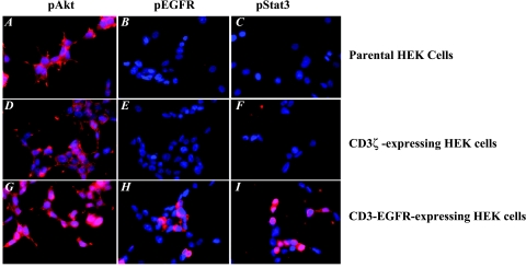
Figure 2.
Parental HEK cells and cells expressing CD3ζ or CD3-EGFR were plated onto four-chamber slides at a density of 2 x 10⁴ cells per chamber in DMEM containing 10% fetal bovine serum. After a 24-hour incubation period, the cells were fixed in acetone and stained with antibodies recognizing phosphorylated forms of Akt (A,D, and G), EGFR (B,E, and H), and Stat3 (C,F and I).

