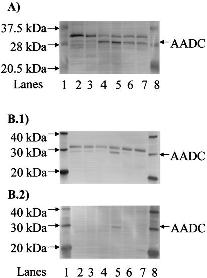
FIG. 3.
Verification of the AADC band in Western blots. (A) Western blot with negative and positive controls for AADC expression. AADC and several of the closest marker bands (20.5, 28, and 37.5 kDa) are indicated. Lanes: 1 and 8, biotinylated protein marker from the horseradish peroxidase protein marker detection pack (New England Biolabs); 2, ATCC 824 at early exponential phase (2-liter bioreactor); 3, strain M5 at stationary phase (static-flask culture); 4, ATCC 824 at stationary phase (5-liter bioreactor); 5, ATCC 824 at transitional phase (5-liter bioreactor); 6, ATCC 824 at stationary phase (2-liter bioreactor); 7, ATCC 824 at transitional phase (2-liter bioreactor). (B) Improved detection of AADC in Western blots. (B.1) Western blot in which antibodies were added in the presence of blocking reagent. (B.2) Western blot in which the primary antibody was pretreated with crude extracts of strain M5 prior to incubation with the membrane. AADC and several of the closest marker bands (20, 30, and 40 kDa) are indicated. Lanes: 1 and 8, MagicMark Western protein standard Invitrogen; 2, ATCC 824(pADC100AS) at transitional phase; 3, ATCC 824(pADC68AS) at transitional phase; 4, ATCC 824(pADC38AS) at transitional phase; 5, ATCC 824(pSOS95del) at transitional phase; 6, strain M5 at stationary phase; 7, strain M5 at exponential phase.

