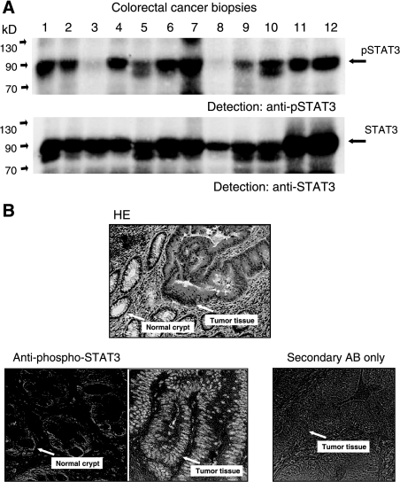
Figure 2.
Analysis of STAT3 tyrosine phosphorylation in colorectal cancer tissue. (A) Examination of lysates from tumor biopsies for activated STAT3 by Western blot analysis. Samples were separated by PAGE, transferred to nitrocellulose, and probed with an antibody specifically recognizing STAT3 phosphorylated on tyrosine 705 (top). Comparable loading of the lanes and identity of STAT3 was confirmed by reprobing the blot with anti-STAT3 (bottom). (B) Histologic examination of a colorectal tumor sample for STAT3 activity. Sections from a representative tumor biopsy were stained with hematoxylin/eosin (top) or immunoreacted with antibody to phospho-STAT3 (bottom). Sections showing differentiated normal cells in crypt structures or dedifferentiated tumor tissues, respectively (arrows), were treated with an antibody specific for STAT3 phosphorylated at tyrosine 705. As a control for antibody and detection specificity, a typical section was stained with peroxidase-coupled secondary antibody without prior treatment with anti-pSTAT3 Tyr 705 (bottom right).

