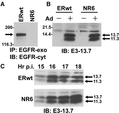
Figure 1.
Characterization of mouse cell lines. (A) ERwt (EGF receptor-positive) and NR6 (EGF receptor-negative) cells were lysed and immunoprecipitated with a human-specific EGF receptor mAb recognizing an exocytic amino acid epitope (EGFR-exo), followed by immunoblotting with an EGF receptor rabbit peptide antiserum to a cytosolic epitope (EGFR-cyt). (B) Cells were mock-treated (−) or infected with adenovirus (+) and harvested 18 h later. (C) Adenovirus-infected cells were harvested 15–18 h after infection (p.i.). (B and C) Total cellular protein (∼80 μg) resolved by SDS-PAGE was immunoblotted with an E3-13.7–specific rabbit peptide antiserum. Molecular weight standards: myosin, 200,000; β-galactosidase, 116,300; soybean trypsin inhibitor, 21,500; lysozyme, 14,400.

