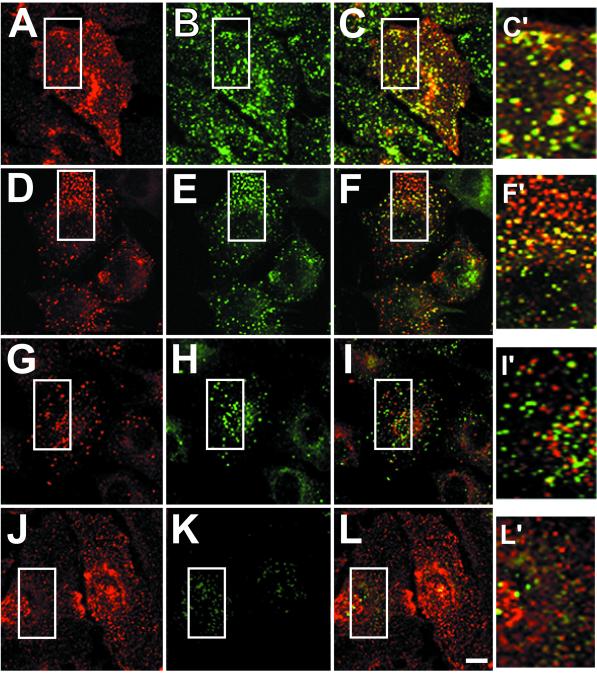
Figure 4.
E3-13.7 colocalization with endocytic marker proteins. Adenovirus-infected A549 cells were permeabilized and fixed at 8 h after infection and then dual-stained for E3-13.7 proteins with a rabbit peptide antiserum and antibodies to transferrin receptor (A–C), Rho B (D–F), Rab 7 (G–I), or cathepsin D (J–L). E3-13.7 is red in A, D, G, and J. Transferrin receptor, Rho B, Rab 7, and cathepsin D are green in B, E, H, and K, respectively. Red and green channels were merged after both fluorescent signals were adjusted to similar levels (C, F, I, and L), and “yellow” indicates the overlap of red and green fluorescence. Insets from merged images are enlarged on the right (C′, F′, I′, and L′). Areas corresponding to the enlarged insets are boxed in all of the panels. All images constitute individual sections from the middle of the cell. Bar, 10 μm.

