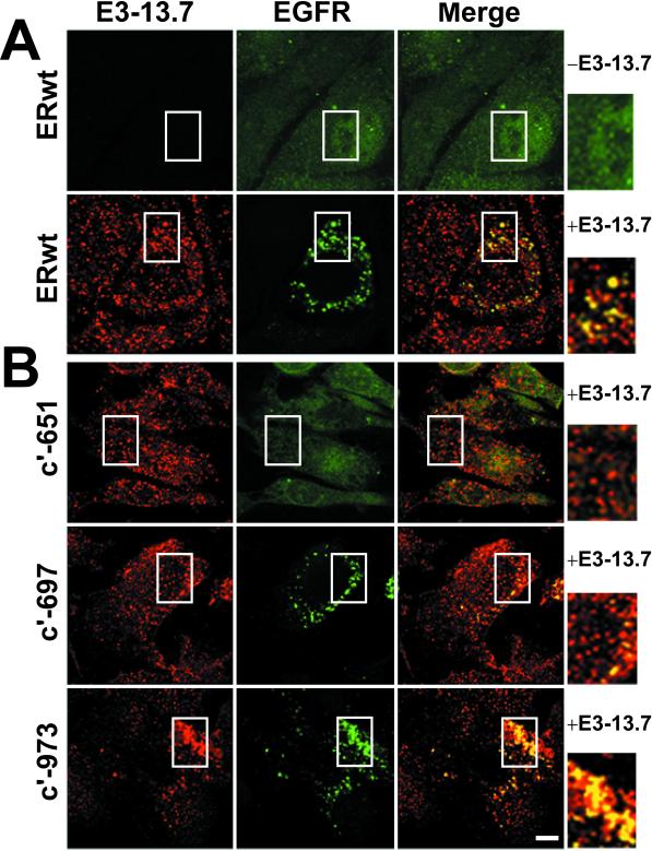
Figure 6.
Intracellular EGF receptors colocalize with E3-13.7. (A) ERwt cells infected with E3-13.7–negative or E3-13.7–positive adenoviruses were stained with a rabbit polyclonal antibody to E3-13.7, a mouse mAb to an external EGF receptor epitope, and corresponding secondary Fab fragments conjugated to rhodamine (donkey anti-rabbit) or fluorescein (goat anti-mouse). Red and green channels were merged after both fluorescent signals were adjusted to similar levels, and “yellow” indicates the overlap of red and green fluorescence. All images constitute individual sections from the middle of the cell. (B) NR6 cell lines expressing cytoplasmically truncated human EGF receptors listed to the left were infected with an E3-13.7–positive adenovirus and then costained for E3-13.7 and EGF receptor exactly as described in A. Insets from merged images are enlarged on the right. Areas corresponding to the enlarged insets are boxed in all of the panels. Bar, 10 μm.

