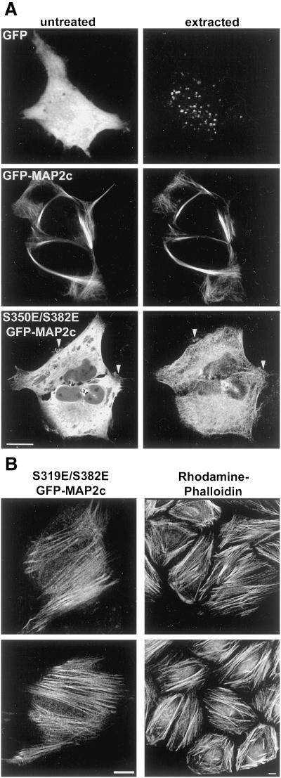
Figure 10.
Specific association between mutant MAP2c and the actin cytoskeleton. (A) Unfixed HeLa cells expressing GFP, GFP-MAP2c, or S350E/S382E GFP-MAP2c were imaged before (untreated) and after extraction (extracted) with a detergent-containing buffer as described in MATERIALS AND METHODS. Virtually all soluble cytoplasmic GFP was removed by this protocol (top). The subcellular distribution and fluorescence intensity of wt GFP-MAP2c was largely unchanged by this procedure (middle). In contrast, the majority of S350E/S382E mutant GFP-MAP2c was extracted (bottom). Labeling was retained in peripheral membrane areas (arrowheads) and along filamentous structures (note thatacquisition parameters were adjusted for display purposes). Bar, 10 μm. (B) After detergent extraction, S319E/S382E GFP-MAP2c was retained in structures resembling actin-based stress fibers, which were prevalent near the bottom of the cell, closest to the glass coverslip. Left, mutant GFP-MAP2c imaged in an unfixed cell after extraction. Right, actin filaments labeled with rhodamine phalloidin in fixed HeLa cells. Bars, 10 μm.

