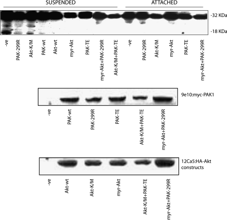
Figure 5.
Cleavage of caspase 3 is inhibited by overexpression of active PAK1 and active Akt. MCF10A cells were transfected with plasmids encoding wild-type (wt), active (PAK-TE or myr-Akt), or dominant-negative (PAK-299R or Akt-K/M) forms of PAK1 and Akt as shown. Cells were grown for 48 hours posttransfection, counted, and split into two plates: one regular culture dish and one polyhema-coated Petri dish for a further 24 hours. Cell lysates were then processed for detection of caspase 3 cleavage, which is revealed by the appearance of an 18-kDa fragment (upper panel). Blots were also probed with 9e10 to detect myc-tagged PAK1 constructs (middle panel) and with 12Ca5 to detect HA-Akt constructs (lower panel).

