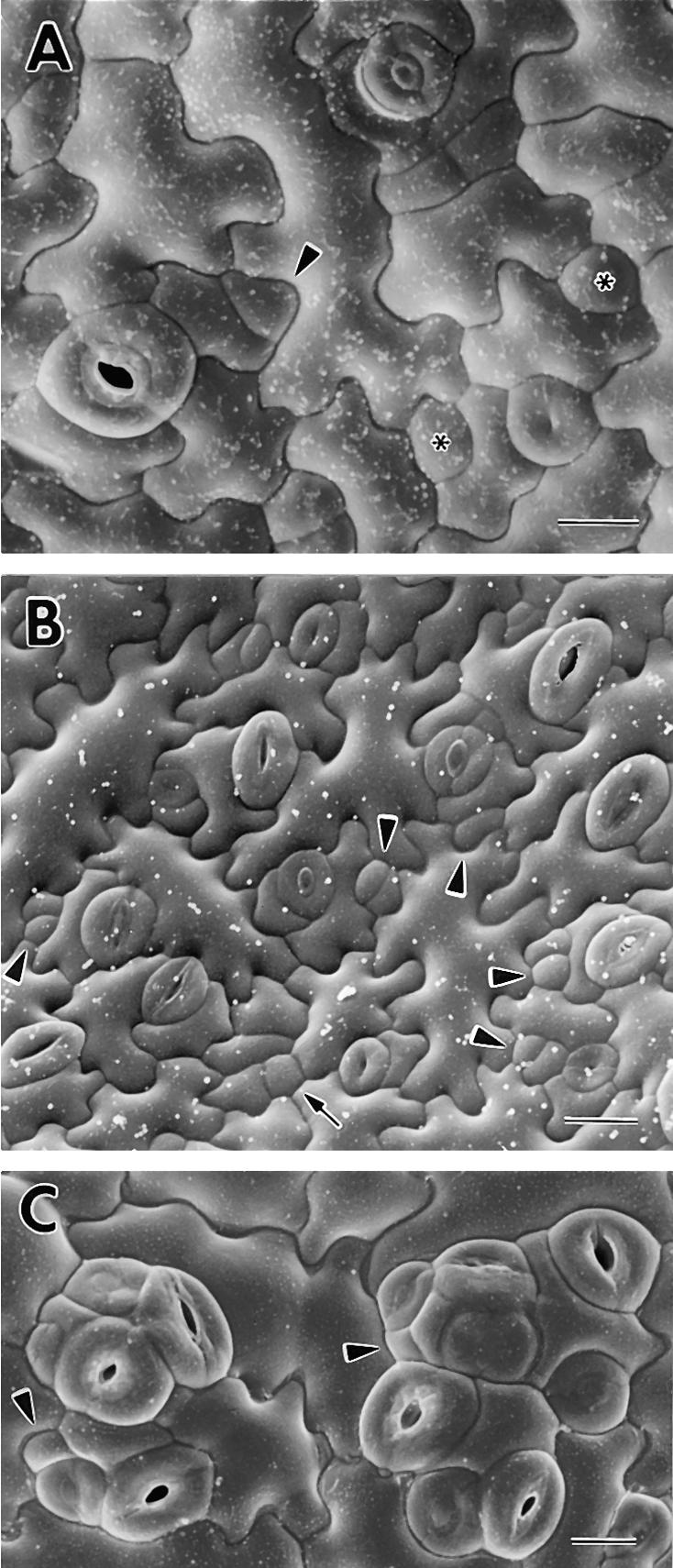
Figure 3.

Cryoscanning Electron Microscopy of Wild-Type Patterning and the Disruption of Stomatal Spacing in tmm.
(A) Wild type. Both the satellite meristemoid (arrowhead) and the larger sister cell (at left) were derived from an asymmetric division of an MMC that was adjacent to a stoma or precursor (now a mature stoma with a pore). Two GMCs (asterisks) were each derived from a satellite meristemoid. Successive stages of pore development are shown at lower and upper right. Note that five of the six existing and future stomata shown are patterned by satellite meristemoid placement.
(B) Wild type. Arrowheads indicate satellite meristemoids, some of which have divided. Arrow shows nonsatellite meristemoid or GMC.
(C) Stomatal clusters in tmm. The arrowheads indicate incorrectly placed satellite meristemoids. Many of the stomata in the clusters are in various stages of pore formation.
 ;
;  .
.
