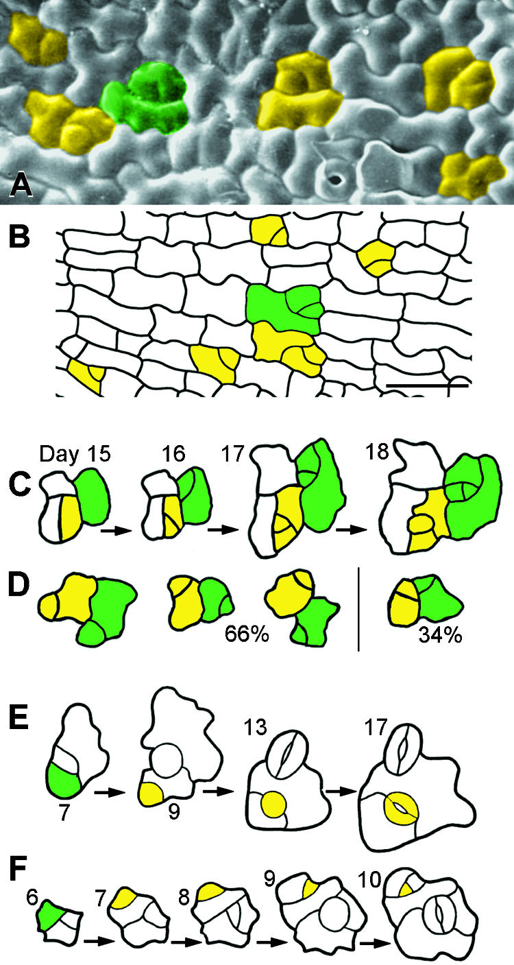
Figure 5.

Placement and Divisions of MMCs.
(A) Scanning electron microscopy of a dental impression from a wild-type cotyledon (4 days after germination), showing that some MMCs are not spaced apart. Each group of cells delimited by color fill came from a single MMC, as deduced from other replicas in the series.
(B) As in (A) except that MMC lineages were deduced from cell wall positions. Shown is a tracing from a fixed cotyledon (12 hr after germination).
(C) Divisions in adjacent MMCs are randomly placed. A dental impression sequence shows divisions of adjacent MMCs that resulted in nonadjacent meristemoids (day 16). Green and yellow areas delimit lineages from each MMC.
(D) As in (C). Sixty-six percent of the divisions of adjacent MMCs yielded separated meristemoids but 34% produced meristemoids in contact. Drawings, which represent different positions, are each from a different dental resin series and summarize results from a sample of fifty adjacent MMCs.
(E) Dental resin series showing that satellite meristemoids are placed away from previously formed precursor cells. The asymmetric division of the MMC (green cell) is oriented so that the new meristemoid (yellow cell at day 9) is separated from the preexisting precursor cell (a meristemoid at day 7; a GMC at day 9) by an intervening sister cell.
(F) As in (E). Both meristemoids divide asymmetrically.
 .
.
