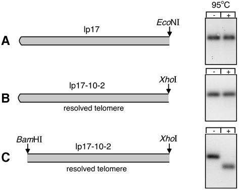
Fig. 5. Test for hairpin telomere at the left end in the resolved substrate. (A) The schematic shows the telomeric structure of wild-type lp17 and the position of an EcoNI site located 2639 bp from the left end. The right panel shows a Southern blot of a 1% agarose gel containing EcoNI-digested B.burgdorferi plasmid DNA hybridized with Probe 1. Duplicate samples were loaded without (–) or with (+) heat treatment at 95°C for 6 min, followed by incubation at 0°C, as indicated. (B) The schematic shows the position of an XhoI site 2538 bp from the left end of the resolved telomere substrate. The blot on the right contains XhoI-digested plasmid DNA from a B.burgdorferi transformant carrying the resolved telomere substrate. The blot was hybridized with Probe 2 (kan). (C) The schematic shows the resolved telomere substrate cut with both XhoI and BamHI to remove the hairpin end on a 70 bp fragment. The blot on the right was also hybridized with Probe 2.

An official website of the United States government
Here's how you know
Official websites use .gov
A
.gov website belongs to an official
government organization in the United States.
Secure .gov websites use HTTPS
A lock (
) or https:// means you've safely
connected to the .gov website. Share sensitive
information only on official, secure websites.
