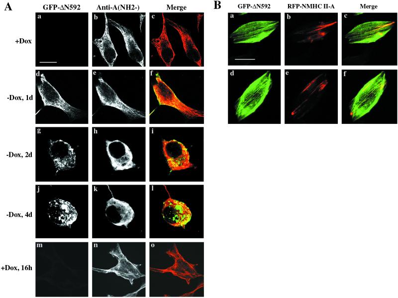
Figure 5.
Disruption of myosin filaments induced by the expression of ΔN592 in HeLa Tet-Off Cells. (A) The cells were cultured in the presence of Dox (a–c) or in the absence of Dox for 1 d (d–f), 2 d (g–i), and 4 d (j–l) or Dox was added after 4 d in culture in the absence of Dox (m–o). An antibody to the NH2 terminus of NMHC II-A was used to distinguish between full-length NMHC II-A (red) and the GFP-ΔN592 fragment (green) in f, i, and l. Yellow indicates colocalization of NMHC II-A and ΔN592. Note cell rounding by 4 d in the absence of Dox (j–l) and the reversal of the phenotype 16 h following addition of Dox (m–o). (B) Colocalization of GFP-ΔN592 and RFP-NMHC II-A. GFP-ΔN592 and RFP-NMHC II-A were transiently cotransfected with L63RhoA into HeLa Tet-Off cells. Transfected cells were fixed with 3.7% paraformaldehyde. GFP-ΔN592 and RFP-NMHC II-A were visualized by green (a and d) and red (b and e) fluorescence, respectively, by using confocal microscopy. The merged images (yellow, c and f) indicate colocalization of GFP-ΔN592 and RFP-NMHC II-A. Images of two different cells are shown. Bar, 20 μm.

