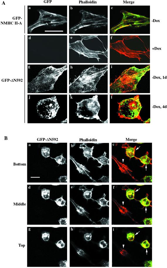
Figure 6.
Rearrangement of actin filaments induced by ΔN592. (A) GFP-NMHC II-A transfected stable cell lines were cultured in the absence of Dox (a–c). Some of the full-length myosin (green) colocalizes with actin (red) to give a yellow image (c). GFP-ΔN592 stable cells were cultured in the presence of Dox (d–f) or in the absence of Dox for 1 d (g–i) or 4 d (j–l). Rearrangement of actin filaments (red) can be seen by day 4 in the absence of Dox. Green indicates ΔN592 and yellow shows areas of overlap between actin (red) and ΔN592 (green). (B) Images of a field after day 4 in the absence of Dox, with two rounded cells (arrows, expressing ΔN592) and one flat cell (arrowhead, not expressing ΔN592) were collected by Z-stack and three different focal planes (bottom, middle, and top) are shown. Bar, 20 μm .

