Figure 3.
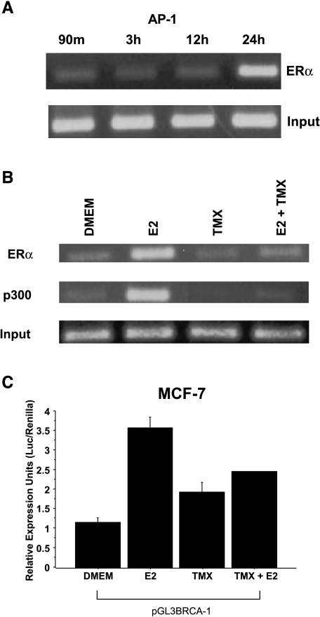
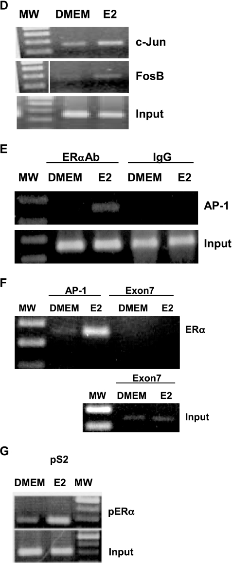
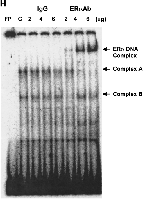
Treatment of MCF-7 cells with estrogen stimulates BRCA-1 promoter occupancy by ERα and p300 to an AP-1 site. (A) MCF-7 cells were precultured for 4 days in DMEM with 5% FBS and then treated with 10 nM E2 for various periods of time. Cells were processed for ChIP assay using an antibody against ERα (Neomarkers, Fremont, CA). Inputs are control bands generated by PCR from cross-linked chromatin. (B) At 24 hours, estrogen stimulates the recruitment of ERα and p300 (antibody from Affinity Bioreagents), whereas 1 µM TMX (Sigma) antagonizes E2-dependent recruitment of ERα and p300. (C) E2-induced BRCA-1 transcription in transiently transfected MCF-7 cells is antagonized by cotreatment with TMX. (D) E2 stimulates the recruitment of c-Jun and FosB. (E) Coincubation with IgG followed by PCR amplification does not produce a band comprising the AP-1 segment. (F) The recruitment of ERα to a region of exon 7 in the BRCA-1 gene (negative control) is not stimulated by E2, which stimulates (G) the recruitment of ERα to an ERE in the pS2 gene (positive control). The size of the amplicon was 237 bp for BRCA-1 (-98 to +139 bp from +1 on exon 1B), 289 bp for the pS2 ERE, and 140 bp for exon 7 of the BRCA-1 gene. (H) EMSA for ERα at the BRCA-1 promoter. MCF-7 cells were precultured for 4 days in DMEM with 5% FBS and then treated for 24 hours with E2. Nuclear extracts were coincubated with a 32P-labeled BRCA-1 oligonucleotide (-40/-19 bp) plus various amounts of mouse IgG or an antibody for ERα. The ERα antibody supershifted a complex (band A) in a dose-dependent manner, thus confirming the presence of ERα at this region; FP, free probe.


