Figure 1.
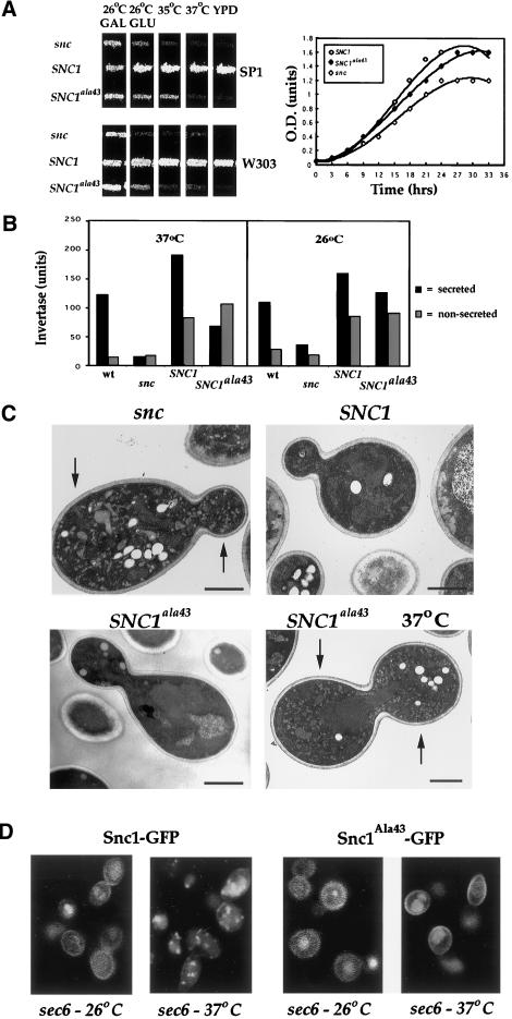
Mutation of methionine-43 to alanine in Snc1 yields a thermosensitive exocytic v-SNARE. (A) SNC1ala43 encodes a thermosensitive v-SNARE. Left panel: snc cells (SP1 background–JG8 and W303 background–JG10) transformed with a control vector (snc) or plasmids constitutively expressing SNC1 or SNC1ala43 were patched onto synthetic complete plates containing galactose. Cells were grown for 3 d at 26°C, before replica plating onto medium containing glucose to deplete SNC1. After 24 h, patches were replica plated onto the following: galactose- (GAL) and glucose-containing (GLU) plates at 26°C; prewarmed glucose-containing plates at 35°C and 37°C; and amino acid-rich medium (YPD). Plates were incubated for 30 h. Right panel: snc cells (JG8) transformed with a control vector (snc), or plasmids expressing SNC1 or SNC1ala43, were grown to log phase on galactose-containing medium and then were shifted to glucose-containing medium for 24 h to deplete SNC1. Cells were seeded in fresh glucose-containing medium at 26°C, and optical density (measured at 600 nm) was monitored as a function of time. (B) Invertase secretion is blocked in SNC1ala43 cells after a shift to restrictive temperatures. Wild-type (SP1), snc (JG8), and snc cells (JG8) transformed with plasmids expressing SNC1 or SNC1ala43 were grown to log phase on galactose-containing medium, shifted to glucose medium for 24 h at 26°C and then derepressed for 2 h on low glucose (0.05%) medium at either 26°C or 37°C. Both secreted (black) and nonsecreted (gray) invertase activities were measured. (C) SNC1ala43 cells accumulate secretory vesicles at restrictive temperatures. snc cells (JG8) expressing a control vector (snc), SNC1, or SNC1ala43 were grown to log phase on glucose-containing medium. Cells were maintained at 26°C, or were shifted for 2 h to 37°C (indicated as 37°C), and were processed for electron microscopy. Bar indicates 1 μm. (D) Localization of Snc1-GFP and Snc1ala43-GFP in sec6 cells. sec6–4 cells transformed with plasmids expressing Snc1-GFP or Snc1ala43-GFP were maintained at 26°C or were shifted for 2 h to 37°C, and then were processed for confocal microscopy.

