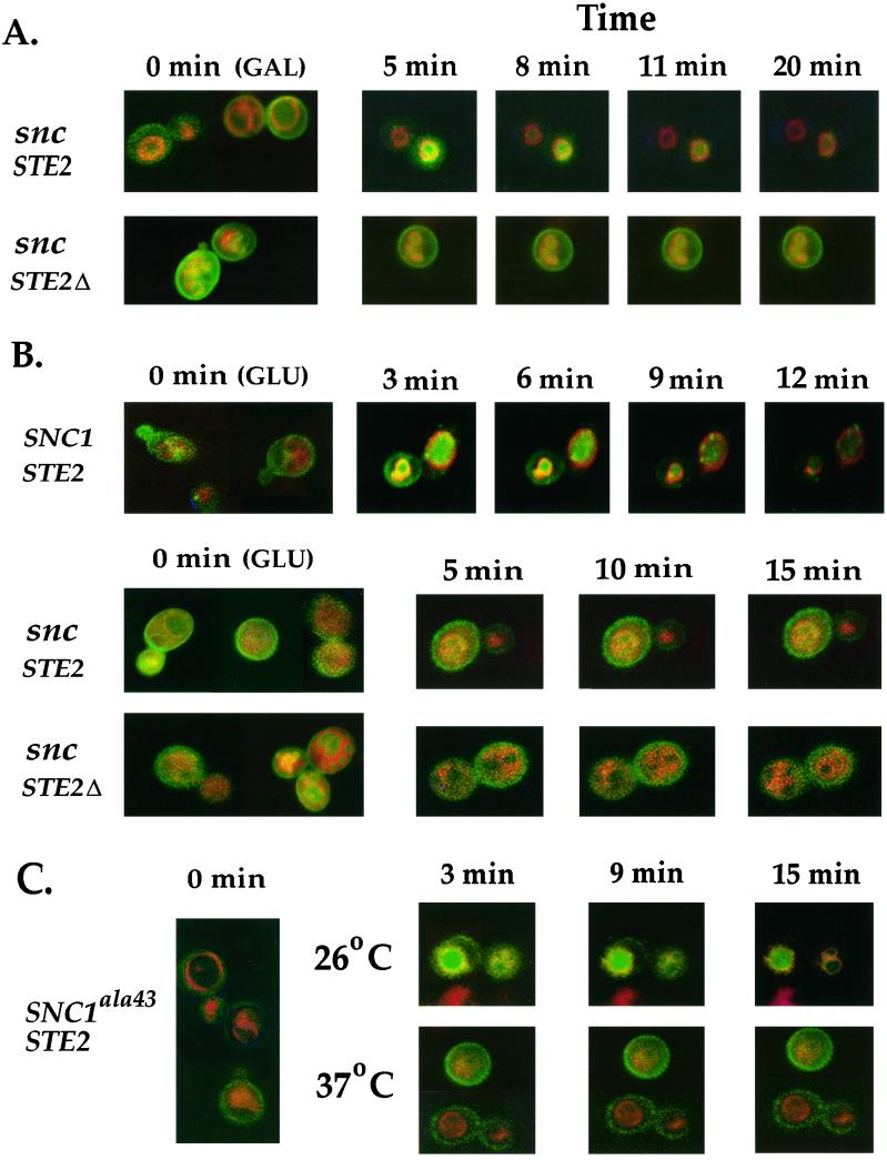
Figure 3.
Ligand-mediated delivery of Ste2 to the vacuole is abolished in snc cells and SNC1ala43 cells shifted to restrictive temperatures. (A) Delivery of Ste2-GFP, but not Ste2Δ-GFP, to the vacuole occurs in snc cells grown on galactose. snc cells (JG8) expressing either Ste2-GFP (snc STE2) or a C-terminal truncated form of Ste2-GFP (snc STE2Δ), which does not undergo endocytosis, were grown to log phase at 26°C before labeling with FM4–64 (see MATERIALS AND METHODS). Cultures were split, whereby half were mounted in soft agar containing no additions, while the other half were mounted in agar containing α-factor (1 μM) and were processed immediately for microscopy. A composite image of untreated cells is representative of the zero time (0 min), while specific α-factor–treated cells were monitored as a function of time. (B) Delivery of Ste2-GFP to the vacuole is abolished in snc cells grown on glucose. snc cells (JG8) expressing SNC1 constitutively or bearing a control plasmid were transformed with plasmids expressing Ste2-GFP to give SNC1 STE2 and snc STE2 strains, respectively. In addition, snc cells expressing the truncated form of Ste2-GFP (snc STE2Δ) also were used. Cells were grown to log phase at 26°C, before labeling with FM4–64. Next, samples of cells were mounted in agar containing no additions, while an equal amount was mounted in agar containing α-factor (1 μM) before visualization, as described above. (C) Delivery of Ste2-GFP to the vacuole is abolished in SNC1ala43 cells shifted to the restrictive temperature. snc cells (JG8) expressing SNC1ala43 were grown to log phase and either incubated with FM4–64 at 26°C or shifted for 30 min to 37°C before labeling with FM4–64 at the restrictive temperature. Cells that were treated with α-factor (1 μM) were monitored over time by microscopy.

