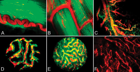
Figure 4.
Imaging of different organs in mouse models. (A) GFP mouse bladder wall imaged using a ×10 regular objective. A typical curled blood vessel (red) embedded into the detrusor muscle fibers (green). (B) A GFP mouse thigh imaged using a ×20 regular objective. Submillimeter vessels (red) are easily identified among skeletal muscle fibers. (C) A Tie2 mouse intact jejunal wall imaged using a ×20 regular objective. Endothelial cells (green) show microcirculation architecture, whereas the intravascular probe (red) reports on blood flow distribution. (D) A Tie2 mouse kidney imaged using a ×20 stick objective through a small flank incision. Individual endothelial cells (green) form the typical glomerular structure of the organ. The intravascular probe (red) is used to show blood flowing through the kidney. (E) A Tie2 mouse liver imaged using a ×20 stick objective. The image is acquired through a small abdominal incision, avoiding manipulation artifacts. (F) A C57BL6/J mouse skull bone marrow imaged using a ×10 regular objective. The intravascular probe (red) can be imaged through intact bones in this region.

