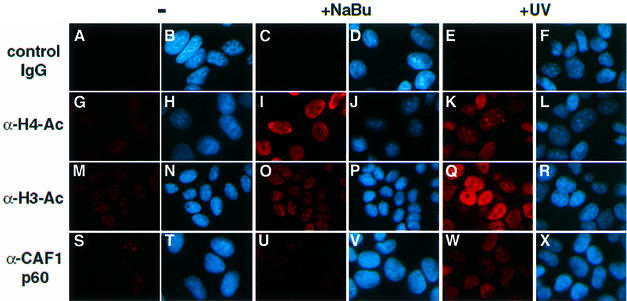
Fig. 6. Histone H3 and H4 acetylation is increased in HeLa cells after UV irradiation. HeLa cells were either untreated (–), UV irradiated (+UV) or treated with sodium butyrate (+NaBu). Cells were then fixed and subjected to immunofluorescence detection. In each column, the right-hand panels show the Hoechst DNA staining (blue), and the left-hand panels correspond to immunodetection with specific antibodies (red), as indicated. A mouse IgG fraction was used as a control in (A), (C) and (E).

An official website of the United States government
Here's how you know
Official websites use .gov
A
.gov website belongs to an official
government organization in the United States.
Secure .gov websites use HTTPS
A lock (
) or https:// means you've safely
connected to the .gov website. Share sensitive
information only on official, secure websites.
