Figure 3.

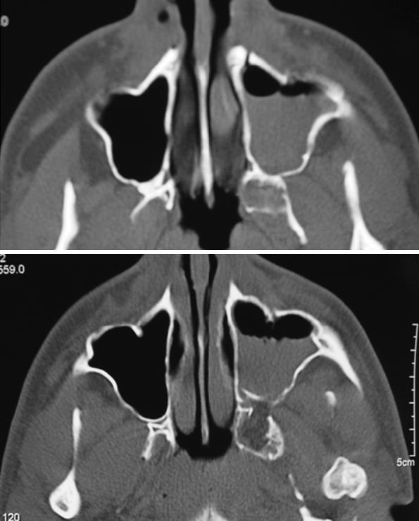
Axial CT 2 days after surgery showing the reposition of the anterior and posterior walls of the left maxillary sinus, the fat obliterating the hole of the removed meningocele, and the postoperative hematosinus. CT, computed tomography.

Axial CT 2 days after surgery showing the reposition of the anterior and posterior walls of the left maxillary sinus, the fat obliterating the hole of the removed meningocele, and the postoperative hematosinus. CT, computed tomography.