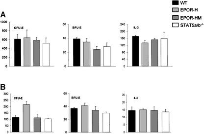
Fig. 4. In vitro colony formation of hematopoietic progenitors from wild-type, EpoR-H, EpoR-HM and Stat5a/b–/– mice in response to various cytokines. The numbers of colonies/105 cells formed from E12.5 fetal liver cell cultures (A) or from bone marrow cell cultures (B) are plotted. The mean and standard deviation are shown from six independent assays. The two-tailed P values for comparison of the various mutant strains with wild-type mice or embryos are >0.01, with the exception of the BFU-E colonies for bone marrow from HM mice (P = 0.004) and of CFU-E colonies for fetal liver cells from H mice (P = 0.00006). Cells from E12.5 embryos or femurs of adult mice were prepared in α-MEM medium containing 2% FBS and counted in the presence of 3% acetic acid to lyse erythrocytes. Diluted cell suspensions and cytokines were mixed with Methocult 3230 to a final concentration of 0.9% methylcellulose. For CFU-E assay, cells were cultured in 0.2 U/ml recombinant hEpo. For BFU-E assay, cells were cultured in 3 U/ml recombinant hEpo and 10 ng/ml recombinant murine IL-3. IL-3 colony assays were performed in the presence of 10 ng/ml recombinant mIL-3. The plating conditions, cell concentration for each assay, culture conditions and colony scoring methods were exactly as described previously (Parganas et al., 1998; Teglund et al., 1998).

An official website of the United States government
Here's how you know
Official websites use .gov
A
.gov website belongs to an official
government organization in the United States.
Secure .gov websites use HTTPS
A lock (
) or https:// means you've safely
connected to the .gov website. Share sensitive
information only on official, secure websites.
