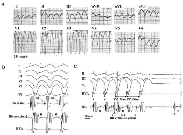
Figure 2.

Twelve-lead ECG during a spontaneous episode of bundle branch reentrant tachycardia (A). Surface ECG leads I, II, III, V1, V2, V6 (B) or II, III, V1 (C) and intracardiac recordings from the His bundle (His) and right ventricular apex (RVA) during bundle branch reentrant tachycardia induced in the same patient. The recordings show many characteristic diagnostic features of bundle branch reentrant tachycardia: (1) typical LBBB morphology and left superior axis (A); (2) AV dissociation (C); (3) H preceding every V with the HV interval (112 ms) greater than that recorded during sinus rhythm (68 ms) (B and C); (5) H precedes the right bundle deflection. This sequence is consistent with ventricular activation through the right bundle branch and is appropriate for a LBBB morphology of tachycardia (B); (6) spontaneous changes in the HH intervals preceded similar changes in the VV intervals (C); and (7) spontaneous termination of tachycardia with retrograde conduction block to H (C). A, H, RB, and V denote atrial, His bundle, right bundle, and ventricular electrograms, respectively. (From: Mazur A, Iakobishvili Z, Kusniec J, Strasberg B. Bundle branch reentrant ventricular tachycardia in a patient with the Brugada electrocardiographic pattern. A.N.E. 2003;8:252-255, with permission of Blackwell Futura Publishing, Inc.)
