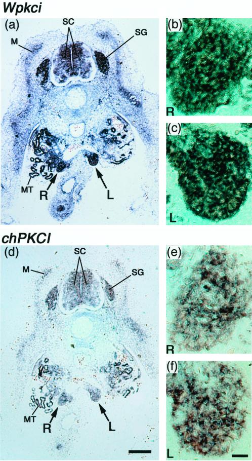
Figure 10.
In situ detection of Wpkci and chPKCI transcripts in the tissue section of 5-d (stages 26 to 27) female chicken embryo. In situ hybridization was carried out as in Figure 9 to the 4-μm-thick paraffin sections containing left (L) and right (R) undifferentiated gonads with the antisense riboprobe for Wpkci (a–c) or chPKCI (d–f). Significant signals of hybridization were detected on both sides of gonads (a and d, and enlarged in b, c, e, and f), mesonephric tubule (MT), spinal cord (SC), spinal ganglion (SG), and myotome (M). Bar in d, 500 μm; bar in f, 50 μm.

