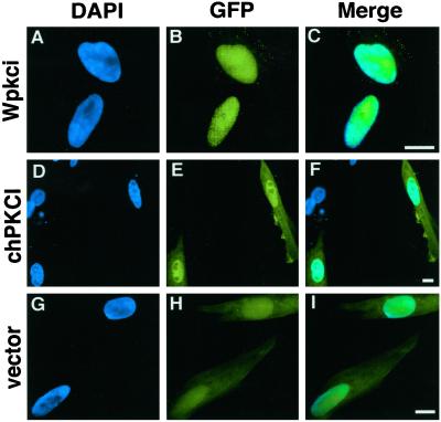
Figure 11.
Intracellular distribution of the GFP-fused form of Wpkci or chPKCI in male chicken embryonic fibroblasts. DAPI-stained nuclei (left panels), GFP fluorescence of the protein expressed (middle panels), and merged images of DAPI staining and GFP fluorescence (right panels) are shown for cells transfected with the expression vector for GFP-Wpkci (A–C), GFP-chPKCI (D–F), or GFP alone (G–I). Bars, 10 μm.

