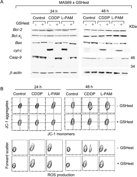
Figure 7.
Ester-mediated GSH increase inhibits the mitochondrial pathway. (A) Immunoblot analysis of cytosolic Bcl-2, Bcl-xl, Bax, cytochrome c, procaspase-9, and β-actin and (B) cytofluorimetric evaluation of Δψm, evaluated by JC-1 staining (upper panel), and ROS production, carried out by DHE probing (bottom panel), performed in both unexposed and GSH ester-exposed MAS69 c-Myc low-expressing transfectant 24 and 48 hours following the end of the treatments with the IC50 doses of CDDP and l-PAM. Each panel is representative of four separate experiments with comparable results.

