Figure 7.
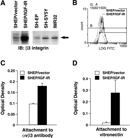
IGF-IR overexpression increases αvβ3 integrin in SHEP NBL cells. (A) SHEP/vector, SHEP/IGF-IR, SHEP, SH-SY5Y, and IMR32 cells were grown to near confluence and whole cell lysates were collected. Lysates were run on a SDS-PAGE gel and Western-immunoblotted for the β3 integrin subunit using a β3 integrin polyclonal antibody. (B) αvβ3 Integrin surface expression measured by flow cytometry. The average relative log fluorescence for SHEP/vector is 17.5 compared with 50.1 for SHEP/IGF-IR. (C) αvβ3 Integrin binding assay. Error bars show SEM (P = .029). (D) SHEP/vector and SHEP/IGF-IR cells were plated on vitronectin. Cells were allowed to attach for 2 hours and rinsed, then adherent cells were stained and read on a fluorimeter [59] (P = .002).

