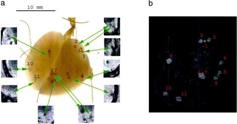
Figure 4.
(a) Photograph of the excised and inflated lungs of a representative mouse with minor tumor load. The tumors were numbered. The cross sections by in vivo micro-CT were indicated on the picture. (b) Semitransparent 3D reconstruction of the same lung with minor tumor load, based on the virtual in vivo CT slices. The spatial organization and localization of the tumor spots can be seen.

