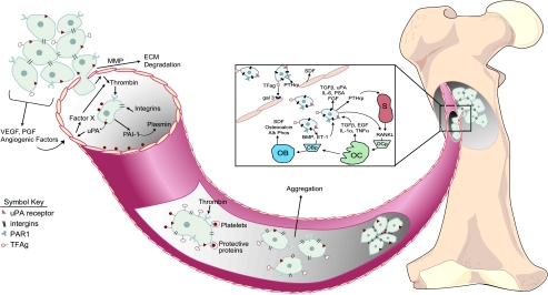
Figure 1.
Diagrammatic representation of cancer metastasis. Initial localized tumorigenesis (first microenvironment) promotes angiogenesis by the release of a variety of angiogenic factors (e.g., vascular endothelial growth factor [VEGF], platelet-derived growth factor [PDGF]). Tumor cells secrete proteases matrix metalloproteinases (MMP) to degrade the extracellular matrices (ECM) and allow the migration of tumor cells into the circulation (third microenvironment). Several factors are released in response to tumor cell intravasation, including urokinase plasminogen activator (uPA), plasminogen activator inhibitor type-1 (PAI1), and thrombin, which promote tumor cell survival and metastasis. The aggregation of tumor cells and platelets during transit promotes survival and ultimately extravasation at the secondary tumor site (second microenvironment). The mechanisms of tumor cell extravasation into the secondary target site of bone are illustrated in the expanded box. Invading tumor cells have been shown to release factors that stimulate both osteoblastic and osteoclastic activity. OBp, osteoblastic progenitor cells; OCp, osteoclastic progenitor cells; OB, osteoblast; OC, osteoclast; S, stromal cell; TFAg, Thomas Friedrich antigen; gal 3, galectin 3 receptor; PTHrp, parathyroid hormone-related protein; SDF, stromal-derived factor.

