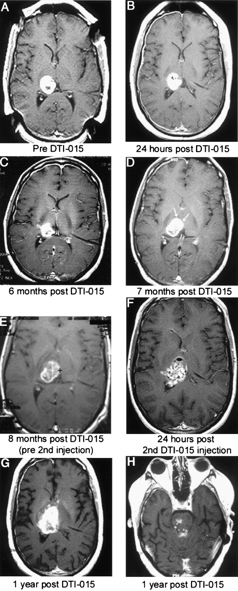
Figure 5.
Patient 6 presented 7 months following diagnosis with a recurrent right thalamic glioblastoma multiforme, which had been increasing in size every 3 weeks following radiation and chemotherapy. All MR images are axial T1-weighted images with contrast. Preinjection MRI tumor volume was 8 ml (A). Twenty-four hours post-DTI-015 (B). Tumor volume decreased to 5.4 ml at 6 months (C). At 7 months, a contrast enhancing area of new tumor growth occurred (D, arrows). A second DTI-015 injection was performed at 8 months (E). Twenty-four hours post-second DTI-015 injection, a mixed hyperintense and hypointense signal is seen (F). One year post-initial DTI-015 treatment, areas of contrast enhancement appeared anterior and superior to the tumor (G) and inferior in the brainstem (H) indicating tumor regrowth. The patient survived 59 weeks from the first DTI-015 injection.

