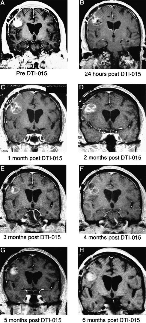
Figure 6.
Patient 29 presented 7 months following diagnosis with a recurrent posterior frontal lobe glioblastoma multiforme following two resections, chemotherapy, and radiation. All MR images are coronal T1 weighted with contrast. Preinjection tumor volume was 3.7 ml (A). Twenty-four hours post-DTI-015 demonstrates a predominantly hypointense signal intensity and decreased contrast enhancement (B). The tumor demonstrated a small ring lesion with central hypointensity at 1–4 months (C–F) and then a slight increase in enhancement at 5 months (G). Six months postinjection (H), the area inferior to the injection progressed as demonstrated by increasing contrast enhancement. At 8 months post-DTI-015 injection, this region and the injected area were resected. The resected region injected with DTI-015 had a “hard rock” consistency. Histological examination of this region demonstrated necrosis, with microscopic foci of tumor at the edges (although no tumor growth). A biopsy of the wall of a previous resection cavity demonstrated tumor mass and diffuse infiltrating tumor.

