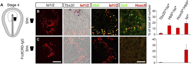
Figure 6. Wnt Signaling Is Required for the Generation of dMNs and vMNs in the Hindbrain and Spinal Cord.
(A) Caudal (C) neural plate tissue explants (black box) were isolated from HH stage 4 embryos. Explants were cultured alone or in the presence of mFrz8CRD-IgG for 28 h and then exposed to Shh-N (15 nM) for an additional 38 h.
(B–C) Bars represent mean ± s.e.m. number of Tbx20 +/Isl +, Hb9 +/Isl +, Hb9 +/Hoxc9 +, and Isl1 + cells, respectively, as percentage of total cell number. Each row represents consecutive sections from a single explant.
(B) Stage 4 C explants cultured with Shh-N alone generated Tbx20 +/Isl + cells in the rostral domain of the explant and Hb9 +/Isl + cells and Hb9 +/Hoxc9 + cells in the caudal domain of the explant ( n = 18 explants).
(C) Explants cultured in the presence of mFrz8CRD-IgG conditioned medium (500 μl/ml culture medium) and Shh-N generated Isl1/2 + cells but no Tbx20 +, Hb9 +, or Hoxc9 + cells ( n = 7 explants). Scale bars represent 100 μm (Isl1/2) and 50 μm (double labels), respectively.

