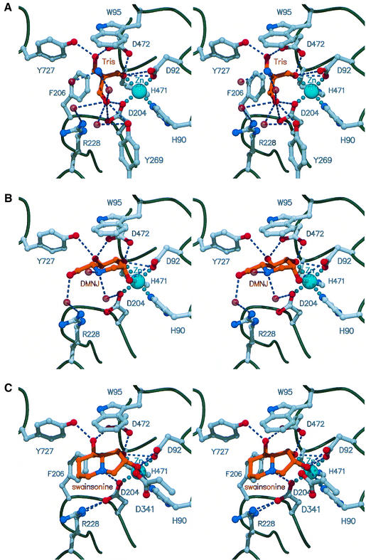
Fig. 3. Stereo views of the active site of dGMII with bound Tris (A), DMNJ (B) and swainsonine (C) molecules. The active site zinc ion is shown in turquoise, the bound inhibitor molecules are rendered in gold and water molecules are represented as transparent red spheres. Interatomic distances <3.2 Å are shown as blue dashed lines.

An official website of the United States government
Here's how you know
Official websites use .gov
A
.gov website belongs to an official
government organization in the United States.
Secure .gov websites use HTTPS
A lock (
) or https:// means you've safely
connected to the .gov website. Share sensitive
information only on official, secure websites.
