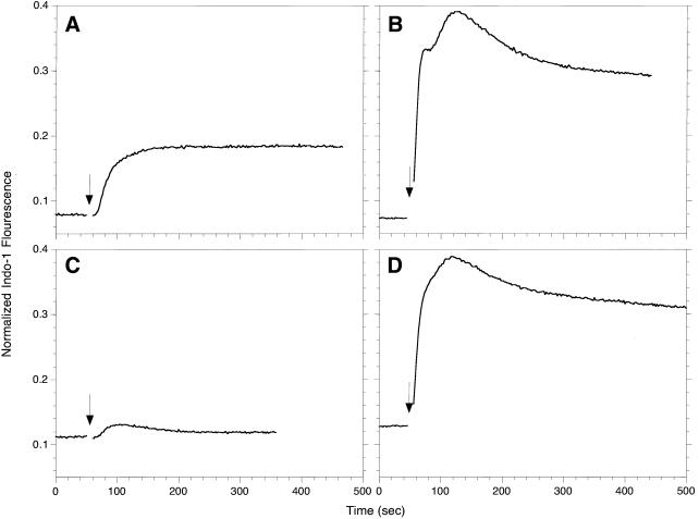
Figure 3.
Ca2+ responses of RBL-2H3 (A, B) or B6A4C1 (C, D) mast cells. IgE sensitized, indo-1 loaded cells were suspended at 106/ml, and indo-1 fluorescence was monitored at 400 nm. Where indicated by an arrow, the cells were stimulated with 100 ng/ml DNP-BSA (A, C) or 250 nM thapsigargin (B, D). The indo-1 fluorescence for each sample was normalized as described in Methods.

