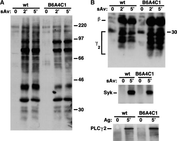
Figure 4.
Antiphosphotyrosine immunoblots of whole cell lysates and immunoprecipitates from RBL-2H3 (wt) and B6A4C1 cells sensitized with biotinylated IgE. (A) Time course of tyrosine phosphorylation stimulated by 10 nM streptavidin for whole cell lysates. (B) Stimulated tyrosine phosphorylation of FcεRI (top), Syk (middle), and PLCγ2 (bottom) immunoprecipitated from cell lysates. Numbers along right margins of immunoblots indicate positions of molecular mass markers in kDa.

