Figure 3.
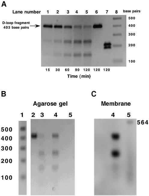
Analysis of endonuclease-mediated cleavage of PCR-amplified mtDNA D-loop fragment. (A) Time course of D-loop fragment cleavage. PCR amplified mtDNA D-loop fragment was incubated in the presence of a 48–64 kDa (see Fig. 1B) active endonuclease fraction (0.05 mg/ml) at 37°C for the times indicated. Aliquots (100 ng) were removed and analyzed by agarose gel electrophoresis. Lanes 1–5, time course; lane 6, mtDNA D-loop incubated for 120 min in the absence of protein; lane 7, SspI cleavage of mtDNA D-loop fragment; lane 8, 1 kb DNA ladder (Life Technologies, Rockville, MA). (B and C) D-loop fragment was amplified using primer 1 5′-end-labeled with fluorescein. Lane 1, 1 kb DNA ladder; lane 2, unlabeled D-loop fragment; lane 3, unlabeled D-loop fragment incubated with active endonuclease; lane 4, labeled D-loop fragment incubated with active endonuclease; lane 5, fluorescein-labeled λ/HindIII fragments. (B) Agarose gel, ethidium bromide stained. (C) DNA transferred to membrane and developed using anti-fluorescein antibody.

