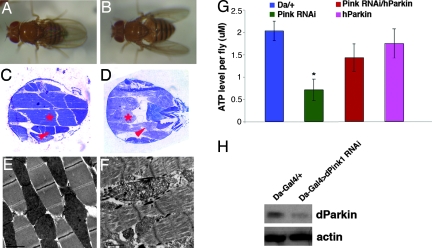
Fig. 5.
Genetic and biochemical interaction between Pink1 and Parkin. Wing posture (A and B), thorax musculature histology (C and D), and IFM EM images (E and F) of Mhc-Gal4/UAS-dPink1 RNAi; UAS-hParkin (A, C, and E), and Mhc-Gal4/UAS-dPink1 RNAi; UAS-hDJ-1 (B, D, and F) flies are shown. Scale bars (2 μm) in E and F are shown at the bottom left corner. (G) Whole-body ATP measurements of the indicated genotypes. ∗, P < 0.01 in Student’s t test. (H) Western blot analysis showing reduction of dParkin levels in dPink1 RNAi animals. Protein extracts prepared from Da-Gal4/+ and Da-Gal4/UAS-dPink1 RNAi animals were probed with anti-dParkin antibody. Actin serves as protein loading control.

