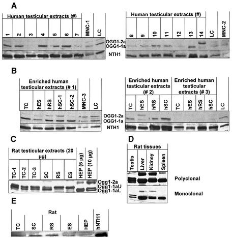
Figure 3.
Western analyses of 8-oxoguanine DNA glycosylase-1 (OGG1) and endonuclease III homologue-1 (NTH1) in human and rat extracts. (A) hOGG1 and hNTH1 in human testicular extracts. Testicular extracts (30 µg/well) were from 14 different testis biopsies, indicated with numbers. Somatic controls were three independent mononuclear blood cell extracts (MNC1-3) and one human lymphoblastoid cell (LC) extract. Upper panels show nuclear hOGG1-1a (lower band) and mitochondrial hOGG1-2a (upper band) using polyclonal anti-hOGG1; lower panels show hNTH1 using polyclonal anti-hNTH1. (B) hOGG1 and hNTH1 in human testicular extracts from enriched spermatogenic cell populations. Cells from three testicular biopsies (nos 1–3) were examined. The spermatocytes (SC) from biopsy 1 were in two different fractions after centrifugal elutriation (hSC-1 and hSC-2). Conditions as in (A). (C) Ogg1 in rat extracts. Extracts from crude and enriched populations of rat male germ cells (20 µg) and from rat primary hepatocytes (5 and 10 µg) were analysed using polyclonal anti-OGG1. Three species are recognised: upper band, Ogg1-2a; middle band, Ogg1-1aU; lower band, Ogg1-1aL. (D) Ogg1 in different rat tissues. Expression of rat Ogg1 in testis, liver, kidney and spleen extracts (20 µg each) analysed with both the polyclonal (upper panel) and the monoclonal (lower panel) OGG1 antibodies. (E) Nth1 in rat extracts. Expression of Nth1 in rat extracts (20 µg each) from different enriched male germ cell populations as well as primary hepatocytes. Abbreviations as in Figure 1.

