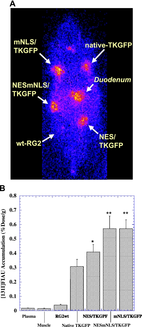
Figure 6.
γ-Camera imaging of [131I]FIAU accumulation in rats bearing multiple subcutaneous tumors produced from RG2 cells expressing native HSV1-tk/GFP or different HSV1-tk/GFP mutants at similar levels based on GFP expression (see dotted cluster box in Figure 5) and from nontransduced RG2 cells (panel A). The levels of [131I]FIAU accumulation measured in tissue samples are expressed as percent injected dose per gram of tissue (panel B). Paired t-test of FIAU accumulation in tumors expressing different mutant HSV1-tk/GFP proteins versus tumors expressing native HSV1-tk/GFP protein (n = 6, *P < .05, **P < .01).

