Fig. 1.
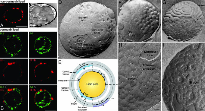
Proteins and structure of the milk fat globule envelope. (A) Immunofluorescence microscopy reveals butyrophilin (But) on the surface of nonpermeabilized milk fat globules. (B) After permeabilization of milk fat globules, adipophilin (Adi), xanthine oxidoreductase (Xor), and TIP47 are detectable. (C) The lipid core of cryosectioned milk fat globules is comprised of many amalgamated small lipid droplet cores. (D) Overview of a freeze-fractured milk fat globule. The globule has been convexly fractured revealing the P-face (PF) of the envelope bilayer with rough areas populated with intramembrane particles (left arrow) and smooth intramembrane particle-poor regions (right arrow). The fracture has penetrated more deeply in some places exposing the underlying E-face equivalent (EFeq) of the envelope monolayer and the concentrically ordered layers of lipid in the core. Entrained cytoplasm is seen between the bilayer and monolayer. (E) Schematic diagram of fracture planes through the milk fat globule. The envelope is comprised of an inner phospholipid monolayer, derived from the cytoplasmic leaflet of the endoplasmatic reticulum membrane, and an outer bilayer, conferred to the milk secretory granule by the plasma membrane during budding, with variable amounts of entrained cytoplasm between. In convex fractures (upper dashed line), the P-face of the bilayer and the E-face equivalent of the monolayer are exposed. In concave fractures (lower dashed line), the E-face (EF) of the bilayer and the P-face of the monolayer are revealed. Asterisks represent sites at which the proteins are labeled with immunogold, and arrows mark the directions from which immunogold labeling in the replicas is viewed. (F–I) Images illustrating variation in morphology of freeze-fractured milk fat globule envelopes. Convex fractures may expose round bumps in the bilayer P-face (F) or elongated bumps with intervening furrows (G). Sometimes, when the fracture penetrates to the underlying monolayer, the E-face equivalent (EFeq) of the monolayer is exposed in planar view underneath the P-face (PF) of the bilayer (H). Intramembrane particles (proteins) in linear arrays are visible within the furrows, and entrained cytoplasm is seen between the bilayer and monolayer. (I) In concave fractures, the E-face (EF) of the bilayer is displayed. Prominent ridges are visible in the E-face of the bilayer. The ridges in the E-face of the bilayer are complementary to the furrows in the P-face of the bilayer. (Scale bars, 5.0 μm in A and B and 0.1 μm in C–I.)

