Fig. 2.
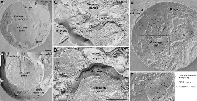
Distribution of adipophilin, TIP47, and xanthine oxidoreductase in freeze-fractured milk fat globules and in mammary epithelial cells. (A) In the globule abundant gold label for adipophilin is seen on the bilayer P-face (PF); the underlying monolayer E-face equivalent (EFeq) is devoid of label. (B) In a mammary epithelial cell, clusters of gold label for adipophilin occur in plasma membrane areas closely apposed to the secretory granule. (B and C) Noticibly lower concentrations of adipophilin label occur in other regions where the plasma membrane is not directly associated with milk secretory granules. The label is located on the P-face of the plasma membrane, i.e., in an equivalent position to the adipophilin label on the globule bilayer P-face. (D) Concave fractures of granules in epithelial cells reveal adipophilin labeling on the monolayer P-face (PF). (B–D) Gold label at the periphery of cross-fractured granules is attributable to adipophilin in the monolayer. No label is present on the bilayer E-face (EF). (E and F) Triple immunogold labeling of adipophilin (18-nm gold), xanthine oxidoreductase (6-nm gold), and TIP47 (12-nm gold) reveals colocalization of these three proteins in the monolayer P-face (PF). Label for all three proteins is absent in the bilayer E-face (EF). (F) A higher magnification view of the boxed area in E. Note the specificity of labeling and complete lack of background. (Scale bars, 0.5 μm in A–D and 0.2 μm in E and F.)

