Fig. 1.
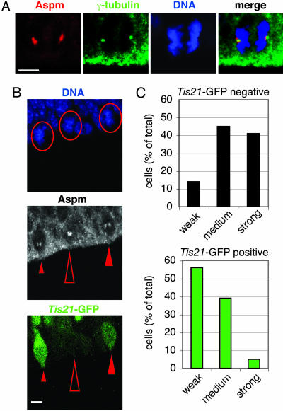
Aspm is concentrated at the spindle poles during mitosis and is down-regulated in NE cells undergoing neurogenic divisions. (A) The dorsal telencephalon of E12.5 Tis21–GFP knockin mice was stained with DAPI (DNA, blue) to reveal NE cells in anaphase and immunostained for Aspm (red) and γ-tubulin (green). All cells analyzed were Tis21–GFP-negative (data not shown). Note the concentration of Aspm in the immediate vicinity of the γ-tubulin-stained centrosomes. (Scale bar, 5 μm.) (B) Comparison of Aspm immunoreactivity at spindle poles in metaphase Tis21–GFP-negative NE cells (proliferative divisions, open arrowheads) versus Tis21–GFP-positive NE cells (neurogenic divisions, filled arrowheads) in the dorsal telencephalon of an E14.5 Tis21–GFP knockin mouse. (Top) DNA staining using DAPI (blue); circles indicate the three metaphase cells analyzed. (Middle) Aspm immunoreactivity (white); the small, medium, and large arrowheads indicate weak, medium, and strong Aspm immunoreactivity at spindle poles, respectively. (Bottom) Tis21–GFP fluorescence (green). (Scale bar, 5 μm.) (A and B) The apical surface of the VZ is down. (C) Quantification in the dorsal telencephalon of E14.5 Tis21–GFP knockin mice of prophase or metaphase Tis21–GFP-negative (black bars, 22 cells) and Tis21–GFP-positive (green bars, 18 cells) NE cells showing weak, medium, or strong Aspm immunoreactivity at spindle poles, expressed as a percentage of total (weak plus medium plus strong). Data are from 19 cryosections that originated from at least four brains.

