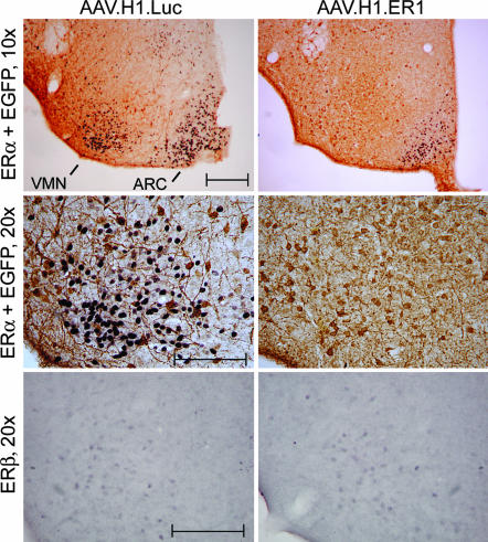
Fig. 2.
AAV-mediated ERα silencing in vivo. Double-label immunostaining for EGFP (brown) and ERα (purple) in the ventromedial area of the hypothalamus of female mice 8 weeks after injections into the VMN with either AAV.H1.Luc or AAV.H1.ER1 (Top). Note a lack of ERα nuclear staining in the VMN but not in the ARC in mice injected with AAV.H1.ER1 compared with a control. Higher-magnification images of the VMN (Middle) reveal that although ERα is not detectable in mice treated with AAV.H1.ER1, the intensity of EGFP staining is comparable in both groups of animals. (Bottom) The number of ERβ-positive cells in the ventrolateral part of the VMH is similar between the two groups, suggesting that AAV.H1.ER1 did not suppress the expression of ERβ. (Scale bar: 200 μm.)

