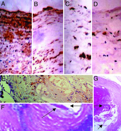
Fig. 2.
Representative immunostaining with the F4/80 antibody in the respective group of mice receiving PE training. (A–D) Lesions from the group receiving the HFD (A) showed extensive staining for macrophage-derived foam cells, but the degree of staining was progressively decreased with MT (B), graduated PE (C), and PE coupled with MT (D). (Magnification, ×400.) (E–G) An example of complex vulnerable aortic plaque (E), higher magnification of a plaque erosion (F) (arrows), and the plaque rupture site (arrow) with an occlusive thrombus (G) in aging mice (16–18 months) receiving HFD. (Magnifications: E, ×400; F, ×1,000; G, ×240.)

