Fig. 4.
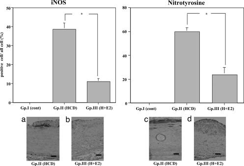
Immunohistochemical analysis with anti-iNOS and anti-nitrotyrosine monoclonal antibody of thoracic aortas of NZW rabbits from atherosclerotic group. (Upper) Area occupied by iNOS-positive cells (Left) and nitrotyrosine-positive cells (Right) in subintimal atherosclerotic plaque of thoracic aortas of rabbits. ∗, P < 0.05. (Lower) Representative photographs of thoracic aortas from rabbits. (Lower Left a) Group II. (Lower Left b) Group III. iNOS was detected adjacent to the necrotic core. (Original magnification, ×100. Scale bars, 25 μm.) (Lower Right c) Group II. (Lower Right d) Group III. Nitrotyrosine was detected in the subintimal atherosclerotic plaque area of the thoracic aortas. (Original magnification, ×100. Scale bars, 25 μm.)

