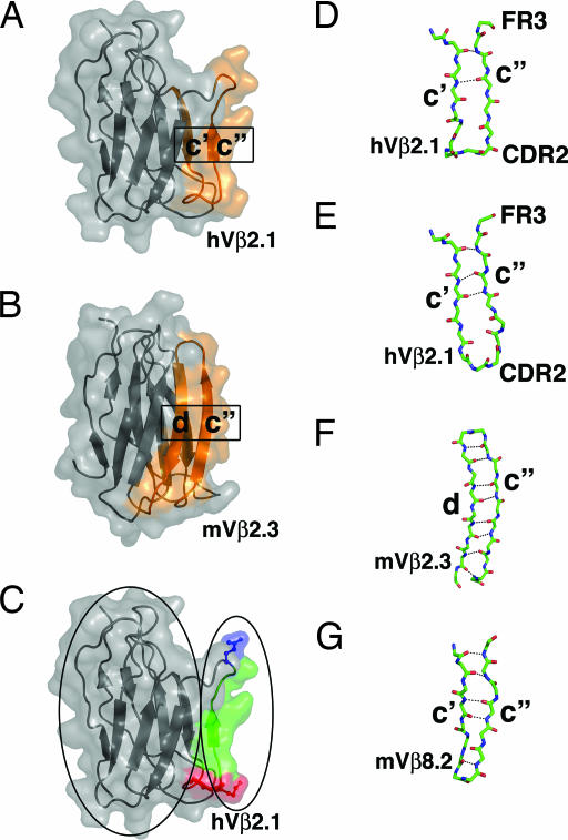
Fig. 3.
A dynamic structural network for the propagation of cooperative binding effects. (A and B) Strand swapping of the c″ β-strand in TCR Vβ domains as depicted in the hVβ2.1 domain (A) (31) and the mVβ2.3 domain (B) (34). The strands that are hydrogen-bonded to one another are colored orange. (C) A view of the hVβ2.1 domain in which the protein core and the CDR2 (red) and FR3 (blue) hot regions and the connecting c″ β-strand (green) are outlined by ovals on the left and right, respectively. (D–G) The c′–c″ or c″–d β-strand regions of hVβ2.1 from a TCR-superantigen structure [Protein Data Bank (PDB) ID code 1KTK] (D), hVβ2.1 from a TCR-autoimmune peptide-MHC structure (PDB ID code 1YMM) (E), mVβ2.3 (PDB ID code 1KB5) (F), and mVβ8.2 (PDB ID code 1BEC) (G).

