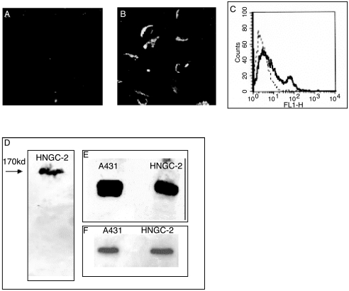
Figure 11.
Expression of c-erbB2 and EGFR. The HNGC-1 cell line was not immunopositive for c-erbB2, as assayed by immunostaining with c-erbB2 antibody (A). About 30% to 40% of the cells from the HNGC-2 cell line showed a strong membranous staining for c-erbB2 (B) and this was quantitatively assayed by flow cytometry wherein, as demonstrated in a representative experiment, a distinct peak of erbB2+ cells is observed with a percent positivity of 35% and an MFI of 38 above a negative cutoff with isotype control of 5% (C). HNGC-2 expressed a 170-kDa EGFR band in Western blotting (D). The expression of c-erbB2 as a 185-kDa protein (E) in the HNGC-2 cell line was comparable to that of the A431 cell line used as a positive control. Equal loading of protein was confirmed by using β-actin loading controls for the two cell lines A431(f) and HNGC-2.

