Figure 1.
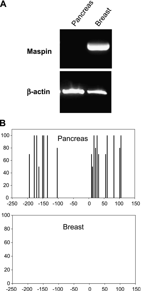
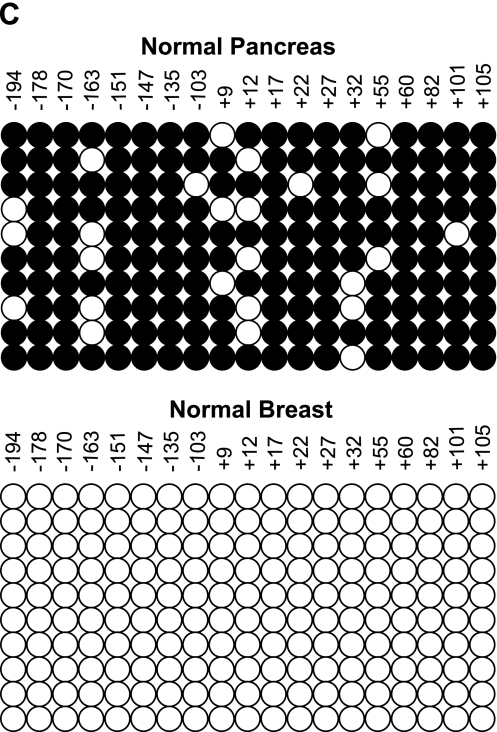
Lack of maspin mRNA in normal pancreatic epithelial cells is tightly associated with promoter methylation status. (A) RNA was isolated from normal pancreas tissue and normal mammary cells (HMECs). RT-PCR was performed for maspin mRNA expression and PCR products were visualized by agarose gel electrophoresis and ethidium bromide staining. Normal pancreas showed no maspin expression whereas normal mammary cells showed robust maspin expression; β-actin was used as a control. (B) DNA from both tissues was sodium bisulfite-modified to determine the methylation status of the maspin promoter. Summary of 5-methylcytosine levels obtained by sodium bisulfite genomic sequencing of the maspin promoter. Cytosine methylation frequency histograms are shown for normal pancreas and HMECs. The x-axis is nucleotide position relative to the transcription start and the y-axis is the percent cytosine methylation. (C) Methylation status of the individual alleles determined by bisulfite sequencing of cloned PCR products. Each row of circles represents the cytosine methylation pattern obtained from individual clones of the maspin promoter. The position of each CpG site relative to transcription start is shown. Open circles indicate unmethylated CpG sites; filled circles indicate methylated CpG sites.


