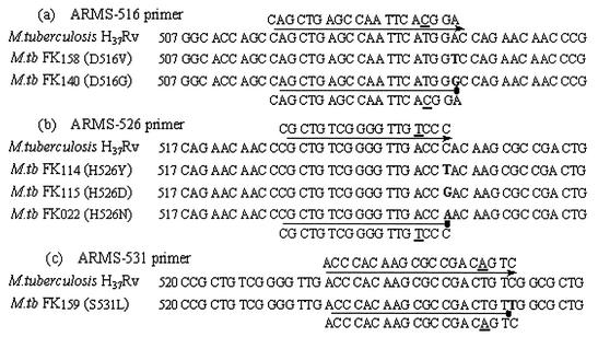
FIG. 1.
Comparison of DNA sequences of rpoB genes in Rifs and Rifr M. tuberculosis isolates. The mutated nucleotides of Rifr isolates are boldfaced. ARMS primers used in this study are shown above and below the sequences. The line with the arrow shows that PCR can be performed well, whereas the line with the dot shows that the ARMS primer is refractory to extension by Taq DNA polymerase. Underlined letters indicate the nucleotide alterations introduced to enhance the 3′ mismatch effect. Codon numbers are assigned on the basis of alignment of the translated E. coli rpoB sequence with a portion of the translated M. tuberculosis sequence and are not the positions of the actual M. tuberculosis rpoB codons.

