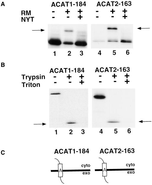
Figure 3.
Orientation of ACAT1-184 and ACAT2-163 in the ER membrane. (A) ACAT1-184 (lanes 1–3) and ACAT2-163 (lanes 4–6) mRNA were translated in vitro in the presence and absence of rough microsomes (RMs) and glycosylation inhibitor (NYT) as described in Figure 2B. Arrows show the position of glycosylated products. (B) Translations of ACAT1-184 and ACAT2-163 were performed in the presence RM and NYT. Reactions were then divided into three equal aliquots and incubated with (+) or without (−) trypsin and Triton X-100. The 14-kDa protected fragments are indicated by arrows. (C) Schematic diagrams of ACAT1-184 and ACAT2-163 exhibiting Ncyt/Cexo membrane topologies.

