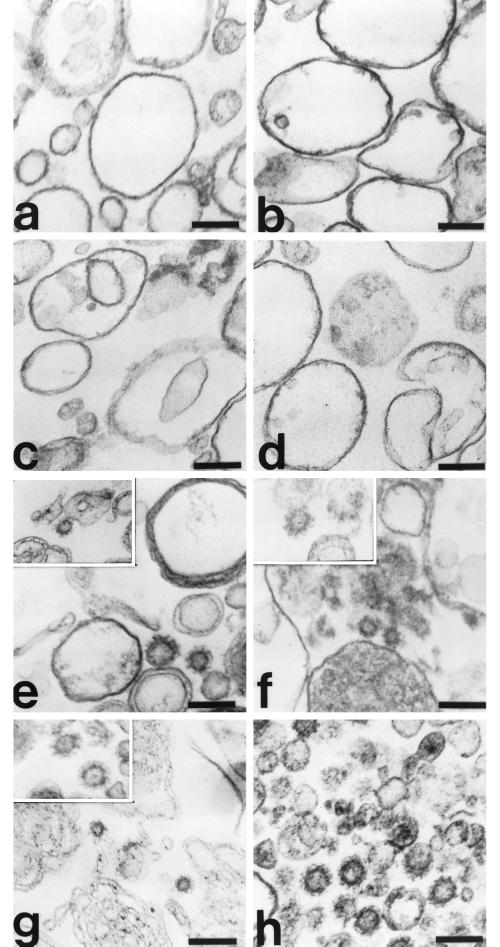
Figure 8.
Morphology of AP-3–containing CCVs assembled on liposomes and Golgi-enriched membranes. Liposomes (200 μg/ml) prepared from soybean 20% PC material (a, c, e, and g) or AP-1– and ARF-depleted Golgi-enriched membranes (50 μg/ml; b, d, and f) were incubated in reactions containing AP-3 (30 μg/ml; a–g), purified clathrin (10 μg/ml; a, b, and e–g), and recombinant myristoylated ARF1 (50 μg/ml; a–g) in the absence (a and b) or presence (c–g) of 100 μM GTPγS at 37°C for 20 min. The membranes were recovered by centrifugation, fixed with 1% glutaraldehyde in 0.1 M sodium cacodylate buffer, pH 7.0, and processed for electron microscopy as detailed in MATERIALS AND METHODS. CCVs isolated from rat liver are shown for comparison (h). Assembly of CCVs in vitro did not occur in the absence of GTPγS (a and b) or in reactions performed in the absence of clathrin (c and d). Bar, 100 nm.

