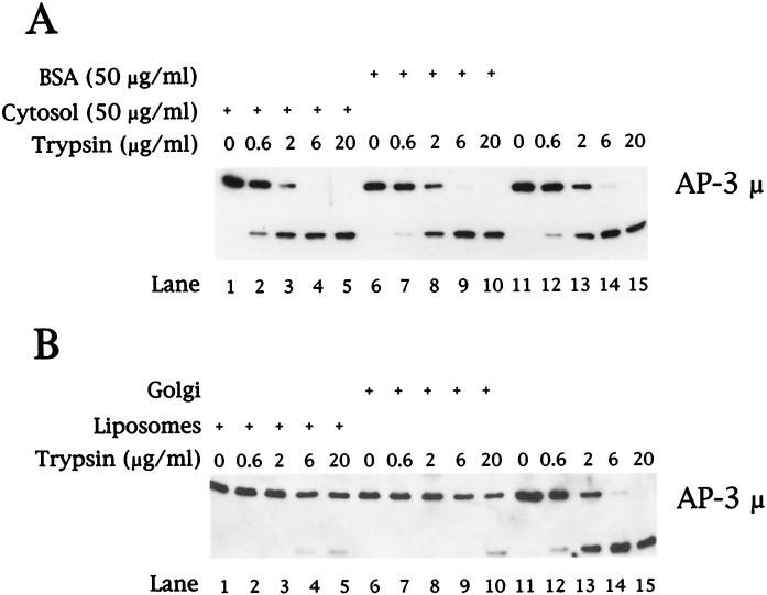
Figure 9.
Controlled tryptic digestion of AP-3. (A) Purified AP-3 (300 ng) was digested with trypsin in the presence of 50 μg/ml bovine adrenal cytosol (lanes 1–5), 50 μg/ml BSA (lanes 6–10), or without exogenous protein (lanes 11–15). After 10 min at 37°C, the reactions were returned to ice and excess soybean trypsin inhibitor was added. Samples were concentrated by methanol/chloroform precipitation, separated by 12% SDS-PAGE followed by transfer to nitrocellulose, and analyzed by immunoblotting with antibodies specific for the μ subunit of the AP-3 complex. (B) Liposomes prepared from soybean 20% PC material (lanes 1–5) or AP-1– and ARF-depleted Golgi-enriched membranes (50 μg/ml; lanes 6–10) were incubated at 37°C for 20 min in reactions containing 5 mg/ml clathrin-depleted bovine adrenal cytosol in the presence of 100 μM GTPγS. Membranes were recovered by centrifugation and resuspended in 1× assay buffer, and equal aliquots were removed for incubation with trypsin as indicated in A. Reactions in which purified AP-3 (300 ng) was digested with trypsin (lanes 11–15) contained 50 μg/ml cytosolic protein.

